D-Dimer
Full Lab Name
Abbreviations
LOINC Code
CPT Code
Methodology
Tube Color
Specimen Type
Minimum Volume
Turnaround Time
Relative Cost
When to Order
- Suspected Deep Vein Thrombosis (DVT) or Pulmonary Embolism (PE) in patients with a LOW or MODERATE pre-test probability (via Wells Score).
- Diagnosis and monitoring of Disseminated Intravascular Coagulation (DIC).
When NOT to Order
- High pre-test probability of PE: Skip the D-Dimer and go straight to CT Pulmonary Angiography (CTPA).
- Post-operative patients, pregnancy, active malignancy, or severe trauma: D-Dimer will be physiologically elevated, rendering the test useless.
EBM Statistics
- Sensitivity: ~95% (Excellent for Ruling OUT disease).
- Specificity: ~40% (Terrible for Ruling IN disease).
- Negative Predictive Value (NPV): > 99% in low-risk patients.
Algorithmic Next Steps
- If < 500 ng/mL: DVT/PE safely ruled out. Stop workup.
- If > 500 ng/mL: DVT/PE NOT ruled out. Proceed to Ultrasound (for DVT) or CTPA (for PE).
Biomarker Kinetics
D-Dimer fragments have a half-life of roughly 8 hours. Levels decline over days following the cessation of thrombosis or initiation of anticoagulation.
Conventional Unit
SI Unit
Conversion Factor
Reference Ranges
| Demographic / Condition | Normal Range |
|---|---|
| Adults (< 50 years) | < 500 ng/mL FEU |
| Adults (> 50 years) | Age × 10 (e.g., 80 years old = < 800 ng/mL) |
Pregnancy Adjustments
D-Dimer naturally increases throughout pregnancy. Standard cutoffs are invalid. Typical median values:
1st Trimester: < 700 ng/mL
2nd Trimester: < 1000 ng/mL
3rd Trimester: < 1700 ng/mL
Critical Values
Physiology
D-Dimer is a specific fibrin degradation product (FDP). It is a small protein fragment present in the blood only after a blood clot has formed and is actively being degraded by plasmin (fibrinolysis).
Causes of Elevation
- Deep Vein Thrombosis (DVT) & Pulmonary Embolism (PE)
- Disseminated Intravascular Coagulation (DIC)
- Recent Surgery or Major Trauma
- Active Malignancy
- Advanced Age & Pregnancy
Causes of Decrease
N/A – Low or undetectable levels represent a normal physiologic state (absence of active thrombosis).
Fasting & Prep
No fasting or special preparation required.
Storage & Transport
Centrifuge within 1 hour to separate plasma. Plasma should be tested within 4 hours if kept at room temperature.
Rejection Criteria
- Underfilled tubes: Alters citrate ratio, causing false results.
- Clotted specimens: In-vitro clotting consumes fibrinogen.
- Severe Hemolysis.
Drug Interferences
High levels of Rheumatoid Factor (RF) can cause false-positive D-Dimer results in some latex agglutination assays.
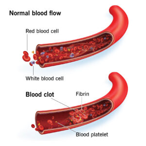
Diagnostic Visuals
Lab Template
Reversal Agents
Direct Oral Anticoagulants (DOACs) and Warfarin do not directly interfere with the D-Dimer ELISA assay itself, though active anticoagulation will naturally lower D-Dimer levels over time by preventing new clot formation.
Pearls & Pitfalls
💡 Pearl: The Age-Adjusted Cutoff
In patients over 50, use the formula (Age × 10). An 80-year-old patient with a D-Dimer of 750 ng/mL has a NORMAL D-Dimer, and PE is ruled out.
Board Focus
- The SNOUT Rule: Highly SeNsitive tests rule OUT disease. Boards love testing that D-Dimer is a rule-out test only.
- PERC Rule: Before ordering a D-Dimer, always apply the Pulmonary Embolism Rule-out Criteria (PERC). If PERC is 0, do not order the D-Dimer.
References
| Citation Title | Link (URL) |
|---|---|
| ACEP Clinical Policy: Suspected Pulmonary Embolism | Link |